Mouse Monoclonal antibody
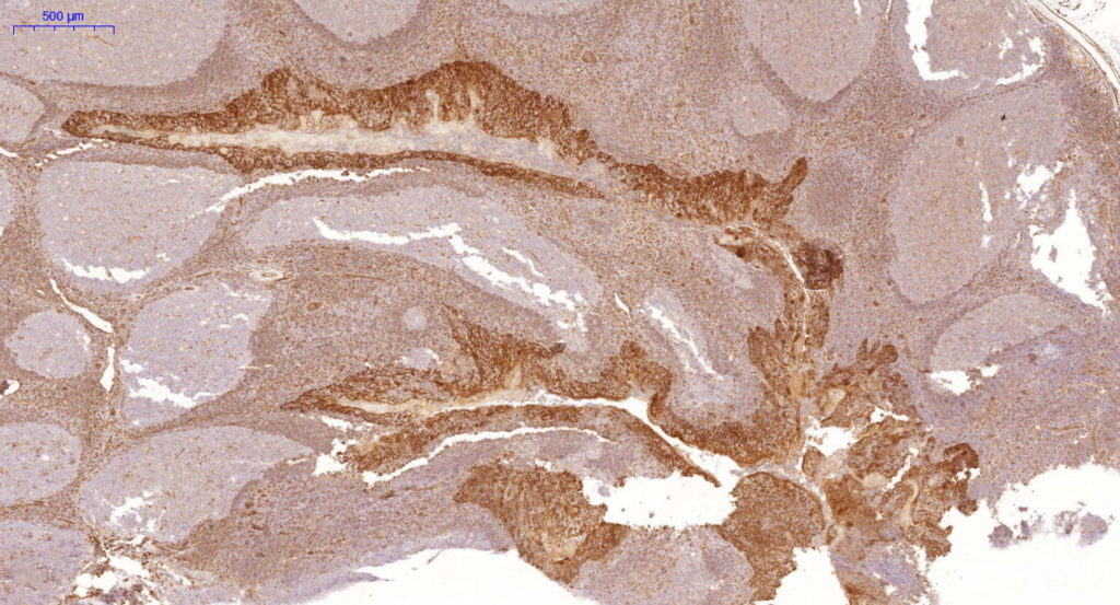
This Mab recognizes a protein of 58kDa, which is identified as Cytokeratin 5 (KRT5). This type II cytokeratin is specifically expressed in the basal layer of the epidermis with family member KRT14. Antibodies to KRT5 identify basal cells of squamous and glandular epithelia, myoepithelial and mesothelium. Anti-Cytokeratin 5 has been reported useful in the differential diagnosis of metastatic carcinoma in the pleura versus epithelioid mesothelioma. Almost all squamous cell carcinomas, half of transitional carcinomas and many undifferentiated large cell carcinomas express. Anti-KRT5, along with anti-p63, affords a high sensitivity and specificity for squamous differentiation. Myoepithelial cells of the breast, glandular epithelia and basal cells of the prostate are labeled with anti-KRT5.
| Catalog No. | Contents | Volume |
| ILM3594-C01 | Cytokeratin 5 | 0,1 ml concentrate |
| ILM3594-C05 | Cytokeratin 5 | 0,5 ml concentrate |
| ILM3594-C1 | Cytokeratin 5 | 1,0 ml concentrate |
Intended use: For Research Use Only
Reactivity: Human, others not known
Clone: KRT5/3594
Species of origin: Mouse
Isotype: IgG1, Kappa
Control tissue: MCF-7 or HeLa cells. Human tonsil , esophagus or bladder tissue.
Staining: Cytoplasmic
Human Entrez Gene ID: 3852
Human SwissProt: P13647
Human Unigene: 433845
Human Chromosome Location: 12q13.13
Immunogen: Recombinant fragment (around aa316-590) of human Cytokeratin 5 (KRT5) protein (exact sequence is proprietary)
Presentation: Bioreactor Concentrate with 0.05% BSA and 0.05% Azide
Application and suggested dilutions:
Pretreatment: Heat induced epitope retrieval in 10 mM citrate buffer, pH6.0 is required for IHC staining on formalin-fixed, paraffin embedded tissue sections. Paraffin embedded tissue section (dilution up to 1:100-1:200)
The optimal dilution for a specific application should be determined by the investigator.
Note: Dilution of the antibody in 10% normal goat serum followed by a goat anti-Mouse secondary antibody-based detection is recommended.
Storage & Stability: Store at 2-8 °C. Do not use after expiration date printed on the vial.
References:
- Ordonez NG. Human Pathology. 2007; 38:1 16.